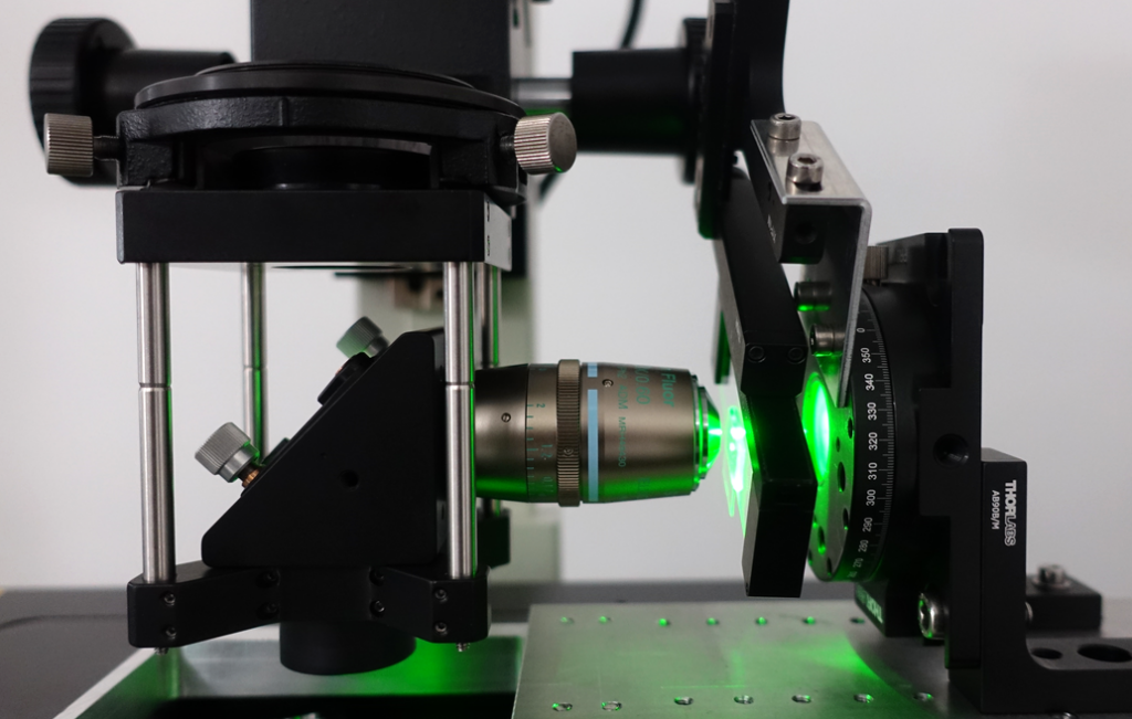
Localisation of Microscopes
FAU OICE works with in-house imaging systems in its purpose-built research building at the IZNF, and with a pool of instruments located at different (de-localised) sites within the FAU.
Terms and conditions for download.
Microscopes can be booked online
- Abberior 3D STED 2-Channel Super Resolution Microscope & Resolft
- Evident Super Resolution Spinning Disc LSM
- iSCAT single molecule imaging Microscope
- LaVision UltraMicroscope II Light Sheet Microscope
- Leica DMi8 TIRF Widefield Fluorescence Microscope & Andor SRRF Camera
- Leica SP5 II Fast Resonant Scanner
- Leica Stellaris 8 Broadband WLL & Tau-Sense LSM
- Leica Thunder 3D Imager Widefield
- Miltenyi UltraMicroscope II Light Sheet Microscope
- Zeiss Celldiscoverer 7
- Zeiss LSM880 NLO Intravital Microscope
- Zeiss Spinning Disc Axio Observer Z1
- Leica SP8 AOBS DIVE Lightning FALCON (AG B. Kost, DFG project number 409476528)
- Zeiss 780 (AG Ch. Alzheimer)
- Leica SP5 II (AG W. Herzog)
- LaVision TriMscope II (AG O. Friedrich)
- STELLARIS 5 upright, with White Light Laser and incubation (AG Boccacini)
- Siemens 7 Tesla MRI (PIPE)
- Brucker ClinScan PET / MRI (PIPE)
- Brucker µCT (PIPE)
- Zeiss Axio Observer 710 (AG Brandstätter)
- Zeiss Axio Examiner 780 (AG Brandstätter)
- Zeiss 710 with Ariyscan (AG F. Engel)
- Leica SP5 II (AG B. Fabry)
- Nikon TiE Epifluorescence Ultrafast-Calcium imaging (AG O. Friedrich)
- Nikon TiE Epifluorescence Ultrafast (AG O. Friedrich)
- Self assembled PALM/STORM Microscope (MPL-MPG) (in-active)
- Nikon TIE (AG J. Winkler)
- Nikon TIE TIRF (AG J. Winkler)